


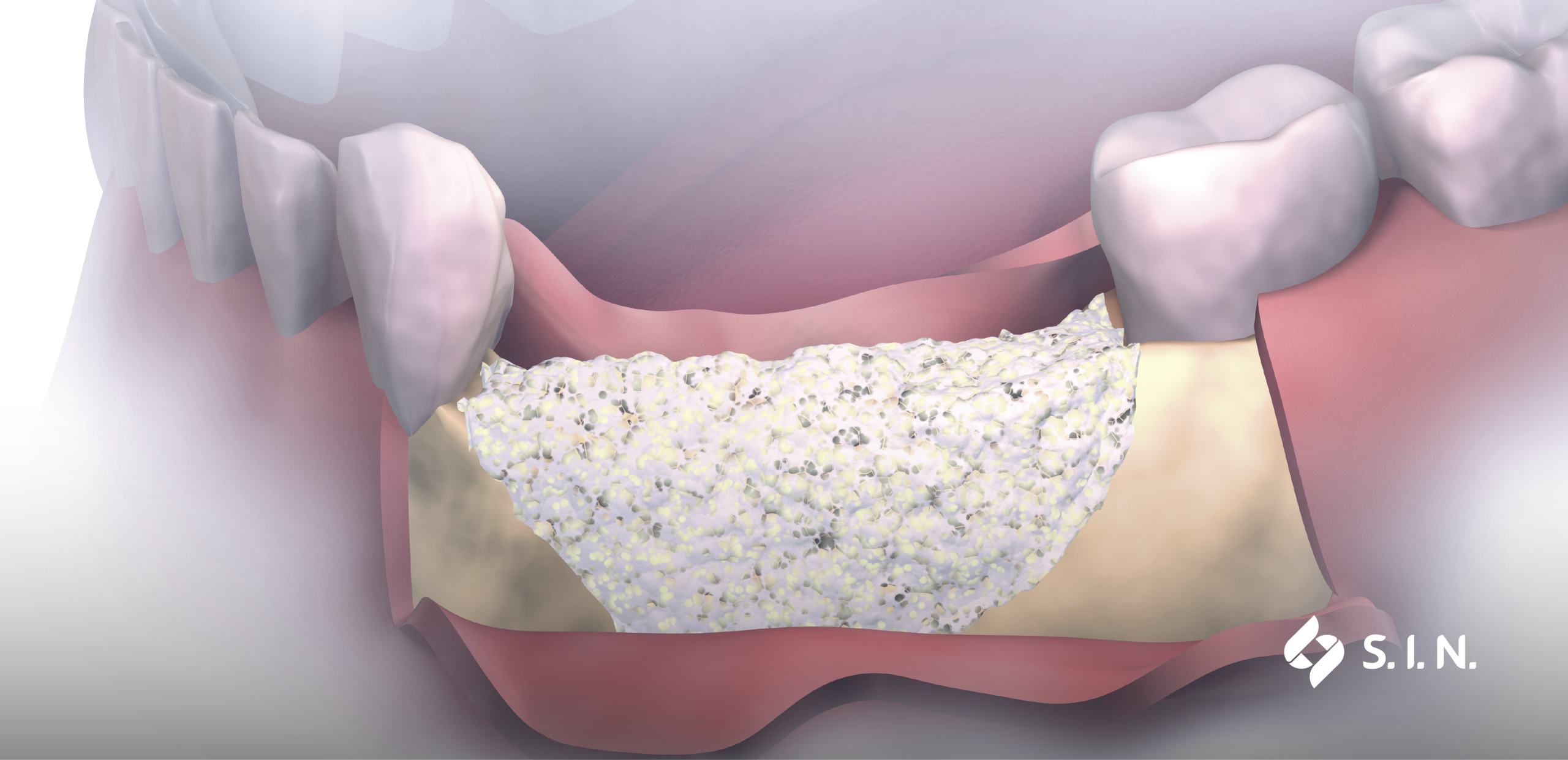
A Dental Surgeon’s technical skills are essential to the success of a treatment, but they aren’t always enough. That’s because an adequate bone structure is imperative to ensure mechanical stability, aesthetics, and longevity for dental implants. In cases of resorption or alveolar ridge defects, Guided Bone Regeneration stands out as one of the most established and predictable techniques in modern dentistry.
Understanding the mechanisms behind this approach is fundamental to structuring safe and efficient treatments. In this article, we delve deep into the biological principles, indications, and materials involved in the success of this technique.
What is Guided Bone Regeneration?
To understand what Guided Bone Regeneration (GBR) is, one must look at the biology of tissue healing. When a bone deficit occurs, different tissues compete to fill that space.
While soft tissues — such as epithelium and connective tissue — gain an early lead in this competition, osteogenic cellular tissue lags behind, which can lead to an imbalance.
This brings us to a clear conclusion: GBR relies on using a physical barrier to isolate the bone defect. By preventing connective tissue cells from invading the site, the membrane creates a protected space so that only osteoblasts and progenitor cells regenerate high-quality bone structure.
Biological and clinical principles of GBR
Guided Bone Regeneration in implantology has well-earned its place, but what leads to its success? It depends on strict adherence to biological and clinical pillars known as the PASS principles:
- Primary closure: the surgical wound must be completely covered by a tension-free flap. Early exposure of the membrane can lead to contamination and procedure failure;
- Angiogenesis: neovascularization is vital to deliver oxygen, nutrients, and osteogenic cells to the graft. Decortication of the recipient bone is frequently performed to stimulate vascularity;
- Space maintenance: the membrane isolates the region, but the space beneath it must be maintained. Combining it with biomaterials and/or physical supports prevents the physical barrier from collapsing under soft tissue compression;
- Stability: the blood clot and graft materials must remain completely immobile. Any micromovement compromises cellular differentiation and new bone formation.

Key indications in implantology
This technique goes far beyond a single objective. Its efficacy is proven and supported by literature and clinicians alike. Highly versatile, GBR can be applied in various clinical scenarios, such as:
- Dehiscences and fenestrations: correction of bone defects around implants placed immediately or in healed sites;
- Alveolar ridge preservation: grafting socket sites immediately after extraction to minimize physiological bone remodeling;
- Ridge augmentation: horizontal or vertical defects in edentulous areas that prevent correct three-dimensional implant placement;
- Peri-implantitis treatment: reconstruction of peri-implant defects resulting from infectious processes.
Materials involved: biomaterials and membranes/barriers
A decisive factor in treatment success is selecting the proper components — and GBR is no exception. Its predictability hinges on choosing the right combination of bone graft material and membrane or barrier.
1. Bone graft substitutes
Options for bone grafting include autogenous, xenogeneic (animal origin), and synthetic materials. While autogenous bone is considered by many to be the gold standard — offering osteogenesis, osteoinduction, and osteoconduction — it requires a secondary donor site and undergoes faster resorption, which can compromise the final regenerative outcome.
Xenogeneic and synthetic grafts, on the other hand, serve as osteoconductive scaffolds. They preserve volume over a longer period thanks to their slower resorption rate.
2. Membranes and barriers
Among the available options, collagen (absorbable) membranes are highly biocompatible and naturally integrate into tissues, eliminating the need for a second surgical procedure for removal. They are ideal for smaller, contained (multi-walled) defects where outcomes are more predictable. These membranes are semi-permeable materials, selectively filtering which cells can pass through the physical obstacle.
In contrast, non-resorbable options — such as e-PTFE, titanium meshes, or customized titanium/zirconia barriers — offer high rigidity and space control. These are indicated for large, complex vertical augmentations, though they require subsequent surgical removal. In other words, they act physically as impermeable barriers, creating complete tissue separation (excluding titanium mesh).
Planning considerations for predictable outcomes
Achieving a predictable aesthetic and functional outcome requires meticulous attention starting in the preoperative phase. Volumetric evaluation using Cone Beam Computed Tomography (CBCT) is indispensable for mapping anatomy and planning the precise quantity of biomaterial needed.
When customized barrier techniques are indicated, intraoral scanning and acquiring virtual Patient models are also essential.
Read also: Intraoral scanning: the equipment transforming the Patient experience
Intraoperatively, delicate soft tissue handling to ensure a tension-free flap, proper instrumentation selection, and firm membrane fixation are critical. Finally, rigorous postoperative monitoring ensures that the bone maturation phase—which typically ranges from 4 to 9 months depending on individual Patient health and case complexity — occurs without unwanted exposure or infectious complications.
Elevate the predictability of your procedures with S.I.N.
Mastering Guided Bone Regeneration requires refined technique, rigorous planning, and the confidence that comes with high-performance biotechnological solutions.
With products developed under strict quality control and backed by scientific evidence, S.I.N. supports your clinical practice from initial planning to the delivery of a new smile — providing the security you and your Patients deserve.

Recapping what we learned in this article:
1. What is Guided Bone Regeneration (GBR) and what is its main goal?
GBR is a surgical technique that uses a physical barrier (membrane/barrier) to isolate a bone defect from fast-healing soft tissues (such as connective tissue). Its primary goal is to protect the space so that only osteoblasts and bone progenitor cells regenerate the bone structure with appropriate volume and quality for dental implant placement.
2. What are the biological pillars of GBR success (PASS Principles)?
GBR success depends on four main pillars known as the PASS principles:
- Primary closure: total wound coverage with a tension-free flap;
- Angiogenesis: promoting neovascularization to supply oxygen and nutrients to the graft;
- Space maintenance: utilizing biomaterials and membranes/barriers to prevent the collapse of the space under soft tissue pressure;
- Stability: complete immobility of the blood clot and biomaterial to allow cellular differentiation and new bone formation.
3. What are the main clinical indications for GBR in Implantology?
GBR is indicated for correcting dehiscences and fenestrations, socket preservation after extraction, horizontal and vertical ridge augmentation in edentulous areas, and reconstructing peri-implant defects caused by peri-implantitis.
4. How do you choose between resorbable and non-resorbable membranes?
The choice depends on the complexity of the bone defect:
- Resorbable membranes (collagen): highly biocompatible and tissue-integrated, eliminating the need for a second surgical removal procedure. Ideal for smaller or contained defects;
- Non-resorbable membranes (e-PTFE, titanium meshes, and zirconia barriers): provide superior rigidity and precise space control, making them ideal for large vertical augmentations and complex reconstructions, though they require later removal.
5. How long does bone maturation take following a GBR procedure?
The maturation time for new bone tissue ranges from 4 to 9 months, depending on defect size, individual Patient physiology and the combination of biomaterials and membranes/barriers used in the treatment plan.